Kerala Plus One Botany Notes Chapter 6 Cell Cycle and Cell Division
Cell cycle:
It involves
- Cell division
- DNA replication
- Cell growth
these all process take place in a coordinated way. The replicated chromosomes (DNA) are then distributed to daughter nuclei.
Phases of Cell Cycle:
Time taken for division:
The duration of cell cycle vary from organism to organism and also from cell type to cell type
- In typical eukaryotic cell cycle (human cells in culture) cells divide once in every 24 hours
- Yeast cell divide in every 90 minutes.
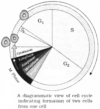
The cell cycle and two basic phases:
- Interphase
- M Phase (Mitosis phase)
Interphase:
The interphase lasts more than 95% of the duration of cell cycle. It is divided into three phases.
1. G1 phase (Gap 1):
G phase is the interval between mitosis and initiation of DNA replication. In this phase cell is metabolically active and continuously grows.
2. S phase (Synthesis):
It is the period which DNA synthesis or replication takes place.
What happens to DNA after S phase?
During S phase amount of DNA per cell doubles. If the initial amount of DNA is denoted as 2C then it Increases to 4C. But the chromosome number is not changed
Events in nucleus and cytoplasm:
In animal cells, during the S phase, DNA replication begins nucleus, and the centriole duplicates in the cytoplasm.
3. G2 phase (Gap 2):
During the G2 phase, proteins are synthesised in preparation for mitosis while cell growth continues.
M Phase (Mitosis phase):
- M Phase represents actual cell division or mitosis
- The M Phase starts with the nuclear division and the separation of daughter chromosomes (karyokinesis).
- It ends with division of cytoplasm (cytokinesis).
Quiescent stage (Go)L
Some cells in the adult animals do not exhibit division (e.g, heart cells), exit G1 phase to enter an inactive stage called quiescent stage.
Common features:
Cells in this stage remain metabolically active but no longer proliferate .But proliferate depending on the requirement of the organism.
M Phase:
This is the most dramatic period of the cell cycle.
Mitosis is an eauational division why?
The number of chromosomes in the parent and progeny cells is the same hence* it is also called as equational division. Mitosis is divided into the following four stages:
- Prophase
- Metaphase
- Anaphase
- Telophase
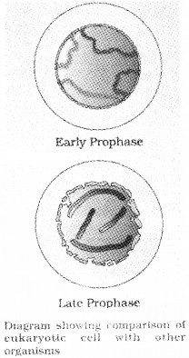
1. Prophase:
It starts after cthe completion of G2 phase.
Key features:
- Chromosomal material condenses to form compact mitotic chromosomes. It consists of two chromatids attached together at the centromere.
- Initiation of the assembly of mitotic spindle fibres.
- At the end of prophase golgi complexes, endoplasmic reticulum, nucleolus and the nuclear envelope disappears.
- The centriole begins to move towards opposite poles of the cell.
2. Metaphase:
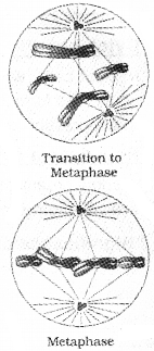
The plane of alignment of the chromosomes at metaphase is referred to as the metaphase plate.
Maximum condensation of chromosome:
In this stage, condensation of chromosomes is completed and morphology of chromosomes can be easily studied. key features:
- Spindle fibres attach to kinetochores of chromosomes.
- Chromosomes are moved to spindle equator and get aligned along metaphase plate through spindle fibres to both poles.
3. Anaphase:
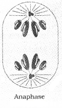
key features:
- Centromeres split and daughter chromatids separate.
- Chromatids move to opposite poles and centromere of each chromosome is towards the pole.
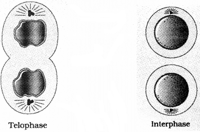
4. Telophase
It is the final stage of mitosis, in which the chromosomes reached their respective poles
key features:
- Chromosomes cluster at opposite spindle poles and their identity is lost as discrete elements. Chromosome decondense as chromatin material.
- Nuclear envelope assembles around the chromosome clusters.
- Nucleolus, golgi complex and ER reappears.
Cytokinesis:
In this two daughter cells separate by a process called cytokinesis.
Cytokinesis in animal cell:
In an animal cell, the appearance of a furrow in the plasma membrane which gradually deepens and ultimately joins in the centre, dividing the cell cytoplasm into two.
Cytokinesis in plant cell:
In plant cells, wall formation starts in the centre of the cell and grows outward to meet the lateral walls. The formation of the new cell wall begins with the formation of a simple precursor, called the cell-plate that represents the middle lamella between the walls of two adjacent cells.
How does a cell become multinucleated?
In some organisms karyokinesis is not followed by cytokinesis as a result of which multinucleate condition arises leading to the formation of syncytium (eg: liquid endosperm in coconut).
Significance of Mitosis:
Mitosis is restricted to the diploid cells only. But in some lower plants and in some social insects haploid cells also divide by mitosis.
- Mitosis results in the production of diploid daughter cells with identical genetic constitution.
- The growth of multicellular organisms is due to mitosis.
- Cell growth results in disturbing the ratio between the nucleus and the cytoplasm.
- Mitosis helps to cell repair, i.e cells of the upper layer of the epidermis, cells of the lining of the gut, and blood cells are being constantly replaced.
- Mitotic divisions in the meristematic tissues – the apical and the lateral cambium, result in a continuous growth of plants throughout their life.
Meiosis:
The cell division that reduces the chromosome number by half results in the production of haploid daughter cells. This kind of division is called meiosis.
What is common to sexually reproducing organisms?
Meiosis ensures the production of haploid phase in the life cycle of sexually reproducing organisms whereas fertilisation restores the diploid phase.
Key features:
- Meiosis involves two sequential cycles of nuclear and cell division called meiosis I and meiosis II but only a single cycle of DNA replication.
- Meiosis I is initiated after the parental chromosomes have replicated to produce identical sister chromatids at the S phase.
- Meiosis involves pairing of homologous chromosomes and recombination between them.
- Four haploid cells are formed at the end of meiosis II.
| Meiosis I | Meiosis II |
| Prophase I | Prophase II |
| Metaphase I | Metaphase II |
| Anaphase I | Anaphase II |
| Telophasel I | Telophasel II |
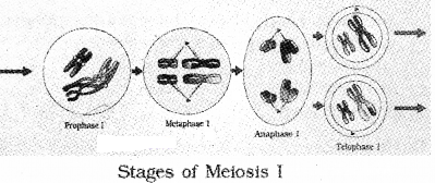
Meiosis I:
Prophase I:
Prophase is typically longer and more complex when compared to prophase of mitosis. It is subdivided into five phases based on chromosomal behaviour i.e., Leptotene, Zygotene, Pachytene, Diploteneand Diakinesis.
| 1. Leptotene stage: The chromosomes become gradually visible under the light microscope. The compaction of chromosomes continues throughout leptotene. |
| 2. Zygotene stage: During this stage homologous chromosomes start pairing together and this process is called synapsis. Synapsis is accompanied by the formation of complex structure called synaptonemal complex. Synapsed homologous chromosome is called a bivalent or a tetrad. The first two stages of prophase I are relatively short-lived. |
| 3. Pachytene stage: During this stage bivalent chromosomes appears as tetrads. This stage is characterised by the appearance of recombination nodules, the sites at which crossing over (exchange of genetic material between two homologous Chromosomes) occurs between non-sister chromatids. The enzyme involved is called recombinase. |
| 4. Diplotene stage: During this stage dissolution of the synaptonemal complex and the tendency chromosomes of the bivalents to separate from each other except at the sites of crossovers. These X-shaped structures, are called chiasmata. In oocytes of some vertebrates, diplotene stage last for months or years |
| 5. Diakinesis stage: During this stage terminalisation of chiasmata occurs. The chromosomes are fully condensed and the meiotic spindle is assembled for separation of chromosomes. By the end of diakinesis, the nucleolus and the nuclear envelope disappears. |
Metaphase I:
The bivalent chromosomes align on the equatorial plate. The spindle fibers attach to the pair of homologous chromosomes.
Anaphase I:
The homologous chromosomes separate, while sister chromatids remain associated at their centromeres.
Telophase I:
The nuclear membrane and nucleolus reappear. After cytokinesis diad of cells are formed. The stage between the two meiotic divisions is called interkinesis. It is short lived. Interkinesis is followed by prophase II, a much simpler prophase than prophase I.
Meiosis II:
Meiosis II resembles a normal mitosis
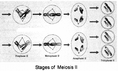
Prophase II:
Meiosis II begins after cytokinesis, The nuclear membrane disappears by the end of prophase II. The chromosomes again become compact.
Metaphase II:
At this stage the chromosomes align at the equator and Spindle fibers get attached to the kinetochores of sister chromatids.
Anaphase II:
It begins with splitting of the centromere of each chromosome allowing them to move toward opposite poles of the cell.
Telophase II:
Meiosis ends with telophase II, in which the two groups of chromosomes get enclosed by a nuclear envelope; cytokinesis follows resulting in the formation of tetrad of cells i.e., four haploid daughter cells.
Significance of meiosis:
| 1. Meiosis conserves the specific chromosome number of each species across generations in sexually reproducing organisms. 2. It results in reduction of chromosome number by half. 3. It increases the genetic variability from one generation to the next. 4. Variations are very important for the process of evolution. |
