Kerala Plus One Botany Notes Chapter 5 Cell The Unit of Life
What is a cell?
Cell is the structural and functional unit of all living organisms. Anton Von Leeuwenhoek first saw and described a living cell Robert Brown discovered the nucleus Unicellular organisms are capable of
- independent existence and
- performing the essential functions of life.
Cell theory:
Schleiden and Schwann together formulated the cell theory:
- In 1838, Malthias Schleiden, a German botanist proposed that all plants are composed of different kinds of cells.
- In 1839 Schwannan British Zoologist, studied different types of animal cells and reported plasma membrane.
Rudolf Virchowd 855) -Contribution of modification of cell theory:
The new cells arise from pre-existing cells (Omnis cellula-e cellula)
Core elements of cell theory:
| (i) All living organisms are composed of cells and products of cells. (ii) All cells arise from pre-existing cells |
An overview of cell:
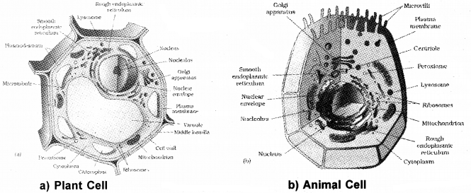
Cell boundary of plant cell and animal cell:
- The onion cell which is a typical plant cell, has a distinct cell wall and inner cell membrane.
- The cells of the human cheek have an outer membrane as the delimiting structure of the cell.
Prokaryotic and eukaryotic cell body:
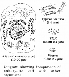
- Cells that have membrane bound nuclei are called eukaryotic whereas cells that lack a membrane bound nucleus are prokaryotic.
- In both prokaryotic and eukaryotic cells, a semi-fluid matrix forms the cytoplasm.
Membrane bound cell organelle of eukaryotes:
- Nucleus
- Endoplasmic reticulum (ER)
- Golgi complex
- Lysosomes
- Mitochondria
- Microbodies
- Vacuoles.
Which is the common cell organelle found in both prokaryotes and eukaryotes?
Ribosomes are non-membrane bound organelles found in both eukaryotic and prokaryotic cell.
- Ribosomes are found not only in the cytoplasm but also within the organelles – chloroplasts and mitochondria and on rough ER.
- Animal cells contain another non-membrane bound organelle called centriole which helps in cell division.
Cells in different measurement:
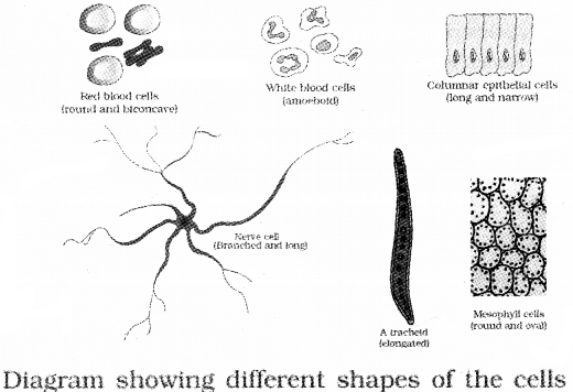
| Mycoplasmas, the smallest cells, are only 0.3μm in length while bacteria is 3 to 5μm Human red blood cells are about 7.0μm in diameter. |
The largest cell is the egg of an ostrich and the longest is Nerve cells.
Prokaryotic cells:
The prokaryotic cells are represented by {bacteria, blue-green algae, mycoplasma and PPLO (Pleuro Pneumonia Like Organisms)}
Classification based on the shape:
- Bacillus (rod like)
- Coccus (spherical
- Vibrium (comma shaped)
- Spirillum (spiral)
(a) The fluid matrix found in the prokaryotic cell is the cytoplasm.
(b) There is no well-defined nucleus
Plasmids:
In addition to the genomic DNA, many bacteria have small circular DNA outside the genomic DNA. These are called plasmids .So they are organisms resistance to antibiotics. The invaginations of plasma membrane seen inside the cell is called mesosome
Cell Envelope and its Modifications:
Three layers of Cell boundary:
- Glycocalyx (Outer)
- The cell wall (Middle)
- Plasma membrane (Inner)
(a) In some bacteria, Glycocalyx is a loose sheath called the slime layer while in others it is thick and tough, called the capsule
(b) Cell wall determines the shape of the cell and provides a strong structural support to prevent the bacterium from bursting.
Mesosome:
They are the extensions of plasma membrane in the form of vesicles, tubules and lamellae.
Functions
They help in
- cell wall formation 1
- DNA replication, distribution.to daughter cells
- respiration
- secretion processes
- increase the surface area of the plasma membrane.
Chromatophores:
Membranous extensions in the cytoplasm which contain pigments. eg: cyanobacteria
Three parts of bacterial flagellum
- Filament
- Hhook
- Basal body.
The other important surface structures in bacteria:
- The pili are elongated tubular structures helps in conjugation
- The fimbriae are small bristle like fibres helps to attach the bacteria on rocks in streams and the host tissues.
Gram +ve and gram -ve:
Christian Gram introduced this method for classifying bacteria. Bacteria that can retain stain(crystal violet) are called Gram positive Bacteria that cannot retain stain are called Gram negative.
Ribosomes and inclusion Bodies:
- In prokaryotes 70S prokaryotic ribosomes consists of subunits – 50S and 30S units.
- Several ribosomes attach to a single mRNA and form a chain called polyribosomes or polysome.
Function:
The ribosomes translate the mRNA into proteins.
Inclusion bodies:
- The examples are phosphate granules, cyanophycean granules and glycogen granules.
- Gas vacuoles are found in blue green and purple and green photosynthetic bacteria.
Eukaryotic cells
They possess well defined and membrance bound cell organelles include
- protists
- plants
- animals
- fungi.
Cell Membrane:
Structure of membrane:
- It consist of lipid bilayer arranged within the membrane with the polar head towards the outer sides and the hydrophobic tails towards the inner part.
- The non polar tail of saturated hydrocarbons is protected from the aqueous environment
- The ratio of protein and lipid varies in different cell types.
- In human beings, the membrane of the erythrocyte has approximately 52 per cent protein and 40 per cent lipids
- The peripheral proteins lie on the surface of membrane while the integral proteins are buried in the membrane.
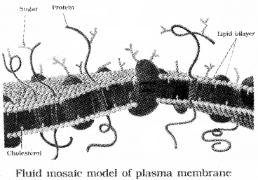
Who proposed the well accepted model of membrane?
Singer and Nicolson (1972) proposed the fluid mosaic model.The quasi-fluid nature of lipid enables lateral movement of proteins within the bilayer.
Functions:
- Transport molecules without energy requirement called as passive transport
- Neutral solutes move across the membrane from higher concentration to the lower by the process of simple diffusion.
- Water move across this membrane from higher to lower concentration by diffusion is called osmosis.
Carrier protein in transport:
As the polar molecules cannot pass through the non polar lipid bilayer, they require a carrier protein to facilitate their transport across the membrane.
Carrier protein and energy in transport:
A few ions or molecules are transported across the membrane from lower to the higher concentration with the help of energy (ATP is utilized). It is called active transport eg: Na+/K+ Pump.
Cell Wall:
Function:
Cell wall gives shape and protects the cell from mechanical damage and infection. It also helps in cell-to-cell interaction and provides barrier to undesirable macromolecules.
Algal cell wall:
It consists Cellulose, galactans, mannans and minerals like calcium carbonate.
Plant cell wall:
It consists of cellulose, hemicellulose, pectins and proteins.
- The cell wall of a young plant cell, the primary wall is capable of growth, which later disappears and secondary wall is formed on the inner (towards membrane) side of the cell
- The middle lamella is made up of calcium pectate which holds the neighbouring cells together.
- Cytoplasmic strands like plasmodesmata which connects cytoplasm of one cell to another through cell wall and middle lamellae.
Endomembrane System:
The endomembrane system include
- endoplasmic reticulum (ER)
- golgicomplex
- lysosomes
- vacuoles.
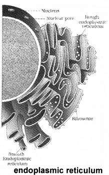
1. The Endoplasmic Reticulum (ER):
Salient features:
- It is the network of tubular structures scattered in the cytoplasm
- ER divides the intracellular space into two distinct compartments, i.e., luminal(inside ER) and extra luminal (cytoplasm)compartments.
Rough endoplasmic reticulum and Smooth endoplasmic reticulum:
The endoplasmic reticulum bearing ribosomes on their surface is called rough endoplasmic reticulum (RER). It is involved in protein synthesis and secretion.
The endoplasmic reticulum devoid of ribosome are called smooth endoplasmic reticulum (SER). It is involved in synthesis of lipids In animal cells, lipid-like steroidal hormones are synthesised
2. Golgi apparatus:
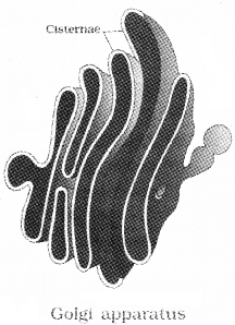
It was first observed Camillo Golgi (1898) as densely stained reticular structures near the nucleus.
Function:
- Packaging of materials
- It is the important site of formation of glycoproteins and glycolipids.
Salient features:
- They consist of many flat, disc-shaped sacs or cisternae of 0.5 μm to 1.0 μm diameter stacked parallel to each other
- The Golgi cisternae are concentrically arranged near the nucleus with distinct convex cis or the forming face and concave trans or the maturing face. The cis and the trans faces are interconnected.
- Materials to be packaged in the form of vesicles from the ER fuse with the cis face of the golgi apparatus and move towards the maturing face.
- The proteins arise from the endoplasmic reticulum are modified in the cisternae of the golgi apparatus and are released from its trans face.
3. Lysosomes:
Salient features:
- * They are membrane bound vesicular structures formed by the process of packaging in the golgi apparatus.
- The hydrolytic enzymes found in these vescicles (hydrolases – lipases, proteases, carbohydrases) are active at the acidic pH.
- These enzymes are capable of digesting carbohydrates, proteins, lipids and nucleic acids.
4. Vacuoles:
Salient features:
- It is the membrane-bound space found in the cytoplasm.
- It contains water, sap, excretory product and other materials
- In plant cells the vacuoles occupy up to 90 percent of the volume of the cell.
- The membrane surrounding the vacuole is the tonoplast,
Function:
It facilitates the transport of ions and other materials against concentration gradients into the vacuole
Type of vacuoles in lower organisms:
In Amoeba the contractile vacuole is important for excretion. In protists, food vacuoles are formed by engulfing the food particles.
Mitochondria:
Salient features:
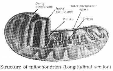
- It is the cylindrical structure having a diameter of 0.2 to 1.0μm
- Each mitochodrion is a double membrane bomd structure.
- The inner compartment is called matrix
- The outer membfrane forms the continous limiting boundary of the oraganelle
- The inner membrane forms a number of infoldings called the cristae that uncreases surface area.
- The matrix possess single circular DNA molecule, a few RNA molecules, and ribosomes(70s)
- The mitochondria divide by fission.
Function:
Mitochondria are the sites of aerobic respiration.
Power house of a cell:
They produce cellular energy in the form of ATP, hence they are called ‘power houses’ of the cell.
Plastids:
Plastids are found in all plant cells and in euglenoids.
Classification of plastids based on the type of pigments:
1. Chloroplasts:
The chloroplasts contain chlorophyll and carotenoid pigments which are responsible for trapping light energy essential for photosynthesis.
2. Chromoplasts:
In the chromoplasts, fat soluble carotenoid pigments like carotene and xanthophylls are present
3. Leucoplasts:
The leucoplasts are the colourless plastids of varied shapes and sizes with stored nutrients:
Classification of leucoplast:
| Amyloplasts store carbohydrates (starch), eg: potato; Elaioplasts store oils and fats Aleuroplasts store proteins |
Chloroplast:
It is found in the mesophyll cells of the leaves. These are lens-shaped,oval, spherical, discoid or even ribbon-like organelles having variable length.
Structure of chloroplast:
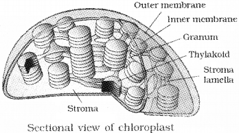
- Chloroplasts are also double membrane bound.
- The space limited by the inner membrane of the chloroplast is called the stroma.
- The stroma contains enzymes required for the synthesis of carbohydrates and proteins.
- It also contains small, double-stranded circular DNA molecules and ribosomes(70S).
- A number of organised flattened membranous sacs called the thylakoids (Chlorophyll pigments seen) are present in the stroma These are arranged in stacks like the piles of coins called grana.
- Stroma lamellae connecting the thylakoids of the different grana.
Ribosomes:
These are granular structures first observed under the electron microscope as dense particles by George Palade(1953).
Chemical composition:
They are composed of ribonucleic acid (RNA) and proteins
Salient features:
- The eukaryotic ribosomes are 80S. Here ‘S’ stands for the sedimentation coefficient
- It consists of two sub units 60S and 40S.
- It translate coded information in mRNA into protiens
Cytoskeleton:
Salient features:
These are network of filamentous proteinaceous structures present in the cytoplasm
Function:
- Mechanical support
- Motility
- Maintenance of the cell shape.
Cilia and Flagella:
Salient features:
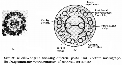
- Cilia and flagella are hair-like outgrowths of the cell membrane..
- Flagella are longer and responsible for cell movement.
- Their core is called the axoneme, possesses a number of microtubules running parallel to the long axis
- The axoneme has nine pairs of doublets of radially arranged peripheral microtubules, and a pair of centrally located microtubules.Such an arrangement is 9 + 2.
Centrosome and Centrioles:
Salient features:
- Centrosome is an organelle containing two cylindrical structures called centrioles
- Both the centrioles in a centrosome lie perpendicular to each other.
- It has cartwheel like organisation and made up of nine peripheral triplet fibrils of tubulin.
- The central part of the centriole is also proteinaceous and called the hub, which is connected with tubules of the peripheral triplets by radial spokes.
Function:
The centrioles form the basal body of cilia or flagella and spindle fibres (give rise to spindle apparatus during cell division in animal cells)
Nucleus:
It was first described by Robert Brown in 1831. Nucleus stained by the basic dyes was given the name chromatin by Flemming
Non nucleated plant and animal cells:
- Erythrocytes of many mammals
- Sieve tube cells of vascular plants
Components of nucleus:
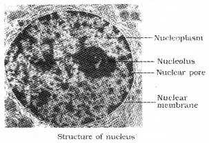
- nucleoplasm
- chromatin
- nuclear matrix
- nucleoli.
Salient Features:
- The outer membrane is continuous with endoplasmic reticulum and bears ribosomes on it.
- These nuclear pores are the passages through which RNA and protein molecules moves.
- The space between two membrane is called the perinuclear space(10 to 50 nm). The nuclear matrix or the nucleoplasm contains nucleolus and chromatin.
- The nucleoli are spherical structures (site for active ribosomal RNA synthesis).
- Larger and numerous nucleoli are present in cells actively carrying out protein synthesis.
- During cell division chromatin condensed to form chromosomes.
Components of chromosome:
- DNA
- basic proteins(histones)
- non-histone proteins
- RNA.
Parts of the chromosome:
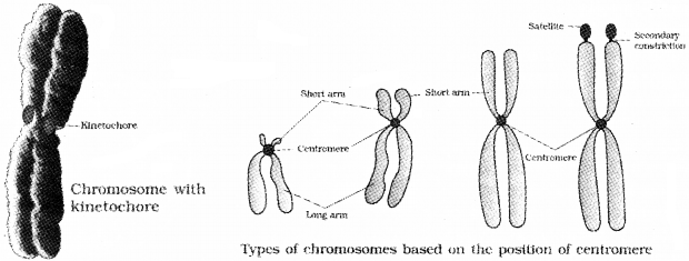
It has primary constriction or the centromere on the sides of which disc shaped structures called kinetochores. A few chromosomes have non-staining secondary constrictions that possess knob like structure called satellite.
Classification of chromosome based on position of centromere:
- Metacentric chromosome has middle centromere forming two equal arms.
- Sub-metacentric chromosome has centromere nearer to one end of the chromosome so it has shorter arm and one longer arm.
- In acrocentric chromosome the centromere is situated close to its end so it has one extremely short and one very long arm.
- Telocentric chromosome has a terminal centromere.
Microbodies:
It is the membrane bound vesicles called microbodies (contain various enzymes) are present in both plant and animal cells.
