ISC Biotechnology Previous Year Question Paper 2015 Solved for Class 12
Maximum Marks: 80
Time allowed: Three hours
- Candidates are allowed additional 15 minutes for only reading the paper. They must NOT start writing during this time.
- Answer Question 1 (Compulsory) from Part I and five questions from Part II, choosing two questions from Section A, two questions from Section B and one question from either Section A or Section B.
- The intended marks for questions or parts of questions are given in brackets [ ].
- Transactions should be recorded in the answer book.
- All calculations should be shown clearly.
- All working, including rough work, should be done on the same page as, and adjacent to the rest of the answer.
Part-1
(Answer all questions)
Question 1.
(a) Mention any one significant difference between each of the following : [5]
(i) Prokaryotic genome and Eukaryotic genome
(ii) Purine and Pyrimidine
(iii) Centrifugation and Crystallography
(iv) Glucose and Glycogen
(v) Codon and Cosmid
(b) Answer the following questions : [5]
(i) Why are Flavr Savor tomatoes preferred over natural tomatoes ?
(ii) State one gene – one enzyme hypothesis.
(iii) Why is the amino acid glycine said to be optically inactive ?
(iv) State Chargaff’s Law of DNA bases.
(v) What is electroporation ?
(c) Write the full form of each of the following: [5]
(i) PIR
(ii) SSB
(iii) STS
(iv) BAC
(v) GBB
(d) Explain briefly: [5]
(i) Insertional inactivation
(ii) Microprocessor
(iii) Supramolecular assembly
(iv) Callus .
(v) Cybrid
Answer:
(a) (i) Prokaryotic genome: In prokaryotic genome, a naked DNA is present, equal to single chromosome.
Eukaryotic genome: In eukaryotic genome, DNA is associated with histone proteins and the number of chromosome is 2 to numerous.
(ii) Purine: They are large size double ring structure e.g., Adenine, Guanine.
Pyrimidine: They are small in size, single ring structures e.g., Thymine, Cytosine.
(iii) Centrifugation: A process to separate small molecules by the action of centrifugal force, simply a physical phenomenon.
Crystallography: It is a technique of studying 3-D structure of macromolecules /atomic arrangement / crystal structure (proteins and nuclear acids) by placing in an intense beam of monochromatic X-rays, producing the regular pattern of reflections.
(iv) Glucose: It is a monosaccharide
Glycogen: It is a polymer formed by condensation of large number of glucose monomers.
(v) Codon: It is a sequence of three nitrogen bases in wRNA which determine the incorpo¬ration of a specific amino acid in a polypeptide chain
Cosmid: They are formed by integration of plasmid with bacterial ori, an antibiotic selection marker, and a cloning site with one or more ‘cos’ sites derived from (λ) lambda bacteriophage.
(b) (i) Flavr Savor tomatoes are genetically modified tomatoes characterised by delayed ripening and longer shelf life.
(ii) One gene-oneenzyme hypothesis is the idea that genes act to the production of enzymes with each gene responsible for producing a single enzyme that intum affects a single step in a metabolic pathway. Concept was proposed by George Beadle and Edward Lawric Tatum in 1941.
(iii) Glycine is optically inactive because it does not affect the plane of polarised light.
(iv) (i) Chargaff’s rule states that purines and pyrimidines are equal in amount ;.e.,A+G = C + T
(ii) Molar amount of purine adenine is always equal to molar amount of thymine. Similarly guanine is equal by cytosine
(iii) Sugar deoxyribosome and phosphate occur in equimolar proportion
(iv) The ratio of A + T/ G + Cis constant for a species.
(v) Electroporation: It is the technique of introducing DNA into the cell by a brief exposure to a very high voltage electric pulse.
(c) (i) PIR – Protein Information Resources
(ii) SSB – Single Strand Breaks
(iii) STS – Sequence Tagged Sites
(iv) BAC – Bacterial Artificial Chromosome
(v) GDB-Genomic Data Bank
(d) (i) Insertional inactivation: Harder problem to solve is to determine which of the transformed colonies comprise cells that contain recombinant DNA molecules, and which contain self-ligated vector molecules. Insertional inactivation is the inactivation of a gene by inserting a fragment of DNA into the middle of its coding sequence. Any future products from the inactivated gene will not work because of the extra codes added to it. Recombinants can therefore, be identified because the characteristic coded by the inactivated gene is no longer visible.
pBR322 contains genes which code for ampicillin resistance and tetracycline resistance. BamHI cuts in the middle of the gene which codes for tetracycline resistance. If a gene is inserted here, the plasmid loses its ability to code for tetracycline resistance. Thus, the plasmid containing the recombinant gene is resistant to ampicillin but sensitive to tetracycline. To screen, we use replica plates.
The pUC8 plasmid is ampicillin resistant and contains a gene lac Z’which partially codes for β galactosidase. To make the plasmid capable of coding for the whole protein, we add the missing DNA along with the recombinant gene. The host which contains the plasmid pUC8 is resistant to ampicillin and is also capable of producing β galactosidase.
(ii) Microprocessor: A microprocessor is a programmable digital electronic component that incorporates the functions of a central processing unit (CPU) on a single integrated circuit (IC).
(iii) Supra molecular assembly is based on weak bonds as supposed to covalent bonds. As mentioned weak bond, though harder to control have the advantage of possible reopening. Relying on weak bond in surface organisation, can lead to molecular assemblies and raise the probability of reorganization processes.
(iv) Callus: It is a mass of meristematic undifferentiated unorganized cells produced in a culture,
(v) Cybrids or a cytoplasmic hybrids are cells or plants containing nucleus of one species but cytoplasm from both the parental species.
Part — II
(Answer any five questions)
Question 2.
(a) Explain the general structure of mRNA and tRNA. Mention their functions during protein
synthesis. [4]
(b) Give one application of each of the following in genetic engineering techniques. [4]
(i) Restriction enzymes and DNA ligases
(ii) Shuttle vectors and Expression vectors
(c) What is meant by genetic code ? [2]
Answer:
(a) Formation of RNA over DNA template is called translation. It is meant for taking the coded information from DNA to the site in cytoplasm where it is required for protein synthesis. Only one of the two strands – sense strand transcribes it. mRNA brings instructions from the DNA for the formation of particular type of polypeptide. The instructions are present in the base sequence of nucleotide’s. Formation of polypeptide occurs over the ribosome, mRNA gets attached to the ribosome.
tRNA are induced to bring amino acids in a particular sequence according to the sequence of codons present over mRNA. mRNA has methylated region at 5′ terminus the cap for attachment with ribosomes. Then followed by initiation code (AUG) either immediately or after a small non-coding region. Then there is coding region followed by termination codon (UAA, UGA, UAG). Then there is non-coding region and poly A’ area at the 3′ terminus. mRNA may be monocistronic (Eukaryote) or polycistronic (Prokaryote).

Transfer RNA (tRNA) : It is also called soluble or sRNA. There are over 100 types of tRNAs. Transfer RNA constitutes about 15% of the total RNA. tRNA is the smallest RNA with 70-85 nucleotides and sedimentation coefficient of 4S. The nitrogen bases of several of its nucleotides get modified, causes coiling of the otherwise single- stranded tRNA into L-shaped form (three-dimensional, Klug, 1974) or clover-like form (two dimensional, Holley, 1965).
It has (i) Anti codon: It is made-up of three nitrogen bases for recognizing and attaching to the codon of mRNA.
(ii) AA-Binding Site : It lies at the 3′ end opposite the anti codon and has CCA—OH group (5′ end bears G). Amino acid or AA-binding site and anti codon are the two recognition sites of tRNA.
Functions : (i) tRNA is adapter molecule which is meant for transferring amino acids to ribosomes for synthesis of polypeptides. There are different tRNA’s for different amino acids. Codons are recognized by anti codons of fRNAs. Specific amino acids are recognized by particular activating or aminoacyl synthetase enzymes.
(ii) They hold peptidyl chains over the mRNAs. The initiator tRNA has the dual function of initiation of protein synthesis as well as bringing in of the first amino acids. There is, however, no tRNA for stop signals.
(b) (i) Restriction enzymes recognize and cleaves a specific short DNA sequence at recognition site. They are used in r-DNA technology as molecular scissors. DNA ligases are the enzymes used to join cut ends of vector DNA and the target DNA by their sticky ends.
(ii) Shuttle vectors is a plasmid containing vector, that can replicate in two different organisms- prokaryote and eukaryote, due to the presence of two different origin of replication i.e, Ori Euk, Ori E-coli.
Expression vectors in addition to incorporate desired gene into the host cell, also help to produce high amount of proteins enclosed by the DNA insert, because of presence of promoter sequence and a terminator sequence along with insert.
(c) Though made-up of only four types of nucleotides, nucleotides of DNA can be positioned in a countless way. As a result, single DNA molecule has several thousand nucleotides, a limitless specificity can be incorporated in the DNA.
There is intimate connection between genes and the synthesis of polypeptides. Genes are made- up of nucleotides, arranged in a specific manner. Arrangement of nucleotides or their nitrogen bases is connected with synthesis of proteins by influencing the corporation of amino acids in them. The relationship between the sequence of amino acids in a polypeptide chain and nucleotide sequence of DNA or mRNA is called genetic code. DNA contains only four types of nucleotide or nitrogen bases while the amino acid is 20. It was, therefore, hypothesized, the triplet code (consisting of three adjacent bases for one amino acid) is operative.
Question 3.
(a) With reference to lipids, explain the following: [4]
(i) Any two chemical properties of lipids.
(ii) Chemical structure and any one function of lipids.
(b) Give the causes and the symptoms of the following metabolic disorders : [4]
(i) Albinism
(ii) Sickle cell anaemia
(c) Name two low resolution techniques used in gene analysis. [2]
Answer:
(a) (i) Chemical properties of Lipids: Hydrolysis: On hydrolysis with alkali or lipolytic enzyme lipases, fats are broken down into their components fatty acids and glycerols.
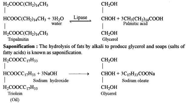
Saponification Value : It is the amount in mg of alkali required to saponify 1 gm of fat or oil. This value is useful for a comparative study of fatty acid chain-length in oils.
(ii) Chemical structure of lipids: Lipids are a heterogeneous group of fats or fat like substances which are insoluble in water but soluble in non-polar solvents like ether, acetone, e.g., ghee, butter, oil, vitamins A, E and K.
Lipids are formed of carbon, hydrogen and often oxygen where oxygen content is very small as compared to other two elements. Lipids require more oxygen for their complete oxidation as compared to carbohydrates. Some lipids also contain small amount of phosphorus nitrogen and rarely sulfur. Most lipids are formed of fatty acids.
Lipids are water insoluble components of the living cells and are essential for storing surplus food. They provide insulation to heat loss and protect internal organs from abrasion. Most biological membranes contain around 50-40% of lipids. Some lipids act as carrier of some vitamins (vitamin A,D, E and K) and also protect them from oxidation. In animals, the fats produce a shock absorbing cushion around eye balls, gonads, kidneys and other vital organs.
(b) (i) Albinism : It is caused by the absence of the enzyme tyrosinase which is essential for the synthesis of the pigment from dihydroxyphenyalanine. The gene for albinism (a) does not produce the enzyme tyrosinase but its normal allele (A) does. Thus, only homozygous individual (aa) is affected by this disease. Albinos (individuals with albinism) lack dark pigment melanin in the skin, hair and iris. Although albinos have poor vision yet they lead normal life.
(ii) Sickle cell anaemia is an autosomal hereditary disorder due to a mutation of single nitrogen base. It results the formation of an abnormal haemoglobin called haemoglobin S (Hbs). In this, only one amino acid-6th amino acid of β-chain glutamic acid is replaced by valine. The erythrocytes become sickle shaped under oxygen deficiency as during strenous exercise and at high altitudes. They cannot pass through narrow capillaries. They clog blood capillaries. Blood circulation and oxygen supply is disturbed. Spleen and brain get damaged. Patient feels acute weakness. Homozygotes (Hbs / Hbs) usually die before reaching maturity.
(c) (i) AFLP genotyping and fingerprinting
(ii) Enzyme electrophoresis
(iii) PCR – SSP technique
Question 4.
(a) With respect to tissue culture techniques, discuss the following : [4]
(i) Various sterilization techniques used.
(ii) Composition of culture medium.
(b ) Discuss the impact of the following factors on enzyme activity : [4]
(i) Substrate concentration and Enzyme concentration
(ii) Temperature and pH
(c) What is single nucleotide polymorphism 9 [2]
Answer:
(a) (i) Various Sterilization Technique :
Maintenance of Aseptic Environment: Dining in vitro culture maintenance of aseptic environment is the most difficult task, because the cultures are easily contaminated by fungi and bacteria present in the air. The contaminants produce toxic metabolites which inhibit growth of cultures plant tissues. Therefore, each step must be handled aseptically and with great care. Following are some of the sterilization methods for aseptic manipulation of plant tissues.
(a) Sterilization of glassware : Glassware (Petri plates, vials, culture tubes, flasks, pipettes, etc.), metallic instruments are sterilized in a hot air oven at 160-180° C for 2-4 hours.
(b) Sterilisation of instruments : The metallic instruments (e.g., forceps, scalpels, needles, spatulas, etc.) are flame sterilised i.e., dipping them in 25% ethanol followed by flaming and cooling. It is called incineration.
(c) Sterilisation of culture room and transfer area: Floor and walls of culture room are washed first with detergent then 2% sodium hypochlorite or 95% ethanol. Larger surface area is sterilised by exposure to UV light. The cabinet of laminar airflow is also sterilised by exposing UV light for 30 minutes and 95% ethanol 15 minutes before beginning of work inside the cabinet of laminar airflow.
(d) Sterilization of nutrient media : Culture media are properly dispensed in glass container, plugged with cotton or sealed with plastic closures and sterilized by autoclaving (steam sterilization) at 15 psi (that gives 121°C) for 30 minutes.
During autoclaving vitamins, plant extracts, amino acids and hormones are denatured. Therefore, the solution of these compounds are sterilized by using millipore filter paper which has 0.2 pm pore diameter.
(e) Sterilization of plant materials : Surface of all plant materials has microbial contaminants. Therefore, disinfectants (e.g., sodium hypochlorite, hydrogen peroxide, mercuric chloride, or ethanol) should be used to make plant materials sterile. Then the chemicals must be washed 6-8 times using sterile distilled water. Then ex plants are transferred aseptically on nutrient medium inside the cabinet of laminar airflow.
(ii) Composition of Nutrient Media: Composition of nutrient media governs the growth and morphogenesis of plant tissue in vitro. Depending on the type of plant cells or tissue used for culture the composition of nutrient media vary. The principal constituents of tissue culture media are inorganic nutrients, carbon sources, organic supplements, growth regulators and gelling agent.
(a) Inorganic nutrients: Several minerals (i.e., macro-and micro-nutrients) are required by the plants. Minerals dissolved in water are dissociated and ionized. For example, in MS medium NH4N03 contributes NO-3 and KNO3 contributes K+ ions. There are six major macro-nutrients such as nitrogen, phosphorus, potassium, calcium, magnesium and sulfur. But the essential or micro-nutrients are required in low amount. These are boron, molybdenum, cobalt, zinc, manganese, iron and copper.
(b) Carbon and energy sources: Mostly sucrose is required as carbon source followed by glucose. These carbon sources enhance cell proliferation and tissue regeneration.
(c) Organic supplements : There is a large number of organic supplements used for best growth of tissues. Vitamins (riboflavin, folic acid ) Amino acids (casein hydrolysate, L-glutamine, L-asperagine, L-glycine, L-arginine, L-cysteine) are commonly used as nitrogen source and enhancers of cell growth. Besides, culture media are also supplemented with casein, coconut milk, yeast and malt extracts, ground banana, orange juice and tomato juice. Activated charcoal added to culture media is known to stimulate plant growth. If required antibiotics (streptomycine or kanamycin) may be added to culture medium to avoid systemic infection of micro-organisms.
(d) Growth regulators : For proliferation of cultured tissues four classes of growth regulators (e.g., auxins, cytokinins, gibberellins and abscisic acid) are used. For induction of root or shoot the ratio of hormones varies considerably. For example, auxins (e.g., indole acetic acid, 1-naphthaleneacetic acid) induce cell division and cause elongation of stem, and intemodes. Cytokinins (e.g., 6-benzylaminopurine, 6-benzyladenine, zeatin) induce cell division and shoot differentiation of cultured tissues. Different ratios of auxins and cytokinins are important in morphogenesis of callus. High ratio of auxin to cytokinin promotes embiyogenesis, callus and root initiation. But high ratio of cytokinin to auxin leads to axillary and shoot promotion.
(e) Solidifying agents : Most commonly agar is used as solidifying or gelling agent. Agar gels do not react with constituents of media and not digested by plant enzymes. Generally 0.5 -1% agar is used to form gel.
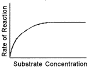
(b) (i) Substrate concentration : It depends on the type of enzyme reaction. For a competitive enzyme reaction, the increase in substrate concentration can have an inverse effect on the activity, but for a normal enzyme reaction, as long as the & substrate concentration isn’t saturated, in general it will increase activity. At lower concentrations, the active sites on most of the enzyme molecules are not filled because there is not much substrate. Higher concentrations cause more-collisions between the molecules. With more molecules and collisions, enzymes are more likely to encounter molecules of reactant. The maximum velocity of a reaction is reached when the active sites are almost continuously filled. Increased substrate concentration after this point will not increase the rate. Reaction rate, therefore, increases as substrate concentration is increased but it levels off.
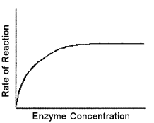
Enzyme Concentration : If there is insufficient enzyme present, the reaction will not proceed as fast as it otherwise would because all of the active sites are occupied with the reaction. Additional active sites could speed up the reaction. As the amount of enzyme is increased, the rate of reaction increases. This is because when more enzyme molecules are present,more substrate molecules can be acted upon at the same time. This means that the total substrate molecules are broken down quickly. If there are more enzyme molecules than are needed, adding additional enzyme will not increase the rate. Reaction rate therefore increases as enzyme concentration increases but then its levels off.
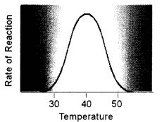
(iii) Temperature : Higher temperature generally causes more collisions among the molecules and therefore, increases the rate of a reaction. More collisions increase the likelihood that substrate will collide with the active site of the enzy me, thus increasing the rate of an enzyme-catalyzed reaction.
Above a certain temperature, activity begins to decline because the enzyme begins to denature. The rate of chemical reactions, therefore, increases with temperature but then decreases as enzymes denature.
Optimum pH : Every enzyme has an optimum pH when it is most effective. A rise or fall in pH reduces enzyme activity by changing the degree of ionization of its side chains. A change in pH may also start reverse reaction. Fumarase catalyses fiimarate → malate at 6.2 pH and reverse at 7.5 pH. Most of the intracellular enzymes function near neutral pH with the exception of several digestive enzymes which work either in acidic range of pH or alkaline, e.g., 2.0 pH for pepsin, 8.5 for trypsin.
(c) Single Nucleotide Polymorphisms (SNPs): SNPs are the variations in a nucleotide at genomic DNA in different individuals at a population which occur due to change even in a single base (e.g.. A, G, T or C). Therefore, certain sites of the sequence nucleotide bases of different
individuals differ as below :
First person — …ATGCTACG…
Second person — …TATCTACG…
In human genome, SNPs occur at 1.6-3.2 million sites. According to changes in bases SNPs affect the gene function. DNA fingerprinting of individuals is possible due to these genetic variations in non-coding parts of genome. This technique is used in search of criminals, rapists, solving parentage problem, confirming identity of individuals, etc.
On average, SNPs occur at every 500-1,000 nucleotides in human DNA, SNPs can help to
- associate sequence variation with heritable phenotypes,
- facilitate studies in population, and
- evolutionary biology, and add in positional cloning and physical mapping.
Question 5.
(a) List the functions of the following bioinformatics tools : [4]
(i) Taxonomy Browser
(ii) BLAST
(iii) ENTREZ
(iv) EMBL
(b) Explain the principle and applications of the following biochemical techniques : [4]
(i) Gel permeation
(ii) Electrophoresis
(c) Give two reasons for germplasm conservation. [2]
Answer:
(a) (i) Taxonomy browser: This search tool provides taxonomic information on various species. The TAXONOMY database of NCBI has information (including scientific and common names) about all organisms for which some sequence information is available (over 79,000 species). The server provides genetic information and the taxonomic relationship of the species in question. TAXONOMY has links with other servers of NCBI e.g., structure and PubMed.
(ii) BLAST (Basic Local Alignment Search Tool) : BLAST is a family of user-friendly sequence similarity search tools on the web. The BLAST server is supported through NCBI (National Center for Biotechnology Information) U.S.A. This tool is designed to identify potential homologous for a given sequence. It can analyse both DNA and protein sequences. A local alignment finds the optimal alignment between subregions or local regions of specified sequences.
(iii) ENTREZ: Integrated information database retrieval system of NCBI. Using Entrez system you can access literature, sequences (both proteins and nucleotides) and structures (3D).
(iv) EMBL (European molecular Biology Laboratory- Nucleic Acid (DNA sequence) databases.
(b) (i) Gel permeation or gel filtration : Size exclusion chromatography (SEC) is a cinematographic method in which particles are separated based on their size, or in more technical terms, their hydrodynamic volume. The name gel permeation chromatography is used when an organic solvent is used as a mobile phase. The main application of gel permeation chromatography is used to analyse the molecular weight distribution of organic-soluble polymer.
Theory and method : The underlying principle of SEC is that particles of different sizes will elute (filter) through a stationary phase at different rates. This results in the separation of a solution of particles based on size, provided that all the particles are loaded simultaneously or near simultaneously.
Particles of the same size should elute together. This is usually achieved with an apparatus called a column which consists of a hollow tube tightly packed with extremely small porous polymer beads designed to have pores of different sizes. These pores may be depressions on the surface or channels through the bead. As the solution travels down the column some particles enter into the pores. Larger particles cannot enter into as many pores. The larger the particles, the less overall volume to traverse over the length of the column and the faster the elution.
The filtered solution that is collected at the end is known as the elute. The void volume includes any particles too large to enter the medium and the solvent volume is known as the column volume.
(ii) Electrophoresis : It is the method of moving charged particles through a medium under the influence on an electric field. It is also used to separate molecules with different physical characteristics using electrical charges.
Types :
- Capillary electrophoresis — commonly used to separate biomolecules by their charge and frictional forces.
- Gel electrophoresis — a technique used by scientists to separate molecules based on physical characteristics such as size, shape, or isoelectric point. It can be used as a preparative technique to partially purify- molecules prior to use of other methods such as mass spectrometry. PCR, cloning, DNA sequencing, or immuno-blotting for further characterization.
Examples of specific techniques include :
- DNA electrophoresis — a specific type of gel electrophoresis used to analyse DNA.
- Protein electrophoresis — a specific type of gel electrophoresis used to analyse proteins.
- Two-dimensional gel electrophoresis — a specific type of gel electrophoresis commonly used to analyse proteins which involves two separation mechanisms to separate molecules.
- SDS PAGE, (sodium dodecyl sulphate polyacrylamide gel electrophoresis) — commonly used to analyse proteins.
(c) Germplasm Conservation : The sum total of all the genes present in a crop and its related species constitutes its germplasm; it is ordinarily represented by a collection of various strains and species. Germplasm provides the raw materials (= genes), which the breeder uses to develop commercial crop varieties. Therefore, germplasm is the basic indispensable ingredient of all breeding programmes, and a great emphasis is placed on collection, evaluation and conservation of germplasm.
Question 6.
(a) Explain the principle and procedure of the PCR technique
(b) Discuss the following innovations in Biotechnology-:
(i) Oil eating bacteria
(ii) Recombinant insulin
(c) What is distant hybridization ?
Answer:
(a) Polymerase chain reaction (PCR) technique was originally invented by Kary Mullis (1985). It results in the selective amplification of a specific region of DNA molecule (up to billion copies). It can generate micro-gram (pg) quantities of DNA copies. The PCR process has been completely automated and compact thermal cycles are available in market.
Principle : The basic principle underlying this technique is that when a double strand DNA molecule is heated to a high temperature, the two DNA strands separate, giving rise to a single stranded DNA molecules (templates). If these single stranded molecules are copied by a DNA polymerase, it would lead to the duplication of original DNA molecule and if these events are repeated many times, multiple copies of the original DNA sequence can be regenerated.
The PCR is carried out in vitro. It utilises the following:
- a DNA preparation containing the desired segment to be amplified (target sequence),
- two nucleotide primers (about 20 bases long) specific, i.e., complementary, to the two 3′-borders (the sequences present at or beyond the 3′-ends of the two strands) of the desired segment,
- the four deoxynucleoside triphosphates, viz. TTP (thymidine triphosphate, dCTP (deoxycytidine triphosphate), dATP (deoxyadenosine triphosphate) and dGTP (deoxyguanosine triphosphate), and
- a heat stable DNA polymerase, e.g., Taq (isolated from the bacterium Thermus acquaticus), Pfu (from Pyrococcus furiosus) and Vent (from Thermococcus litoralis) polymerases. Pfu and Vent polymerases are more efficient than the Taq polymerase.
Procedure of PCR:
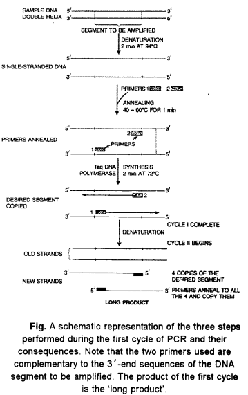
At the start of PCR, the DNA from which a segment is to be amplified, an excess of the two primer molecules, the four deoxyriboside triphosphates and the DNA polymerase are mixed together in the reaction mixture that has appropriate quantities of Mg2+. The following operations are now performed sequentially (Fig.)
Denaturation:
The reaction mixture is first heated to a temperature between 90-98°C (commonly 94°C) that ensures DNA denaturation. This is the denaturation step. The duration of this step in the first cycle of PCR is usually 2 min at 94°C.
Annealing:
The mixture is now cooled to a temperature (generally 40-60°C) that permits annealing of the primer to the complementary sequences in the DNA; these sequences are located at the 3′-ends of the two strands of the desired segment. This step is called annealing. The duration of annealing step is usually 1 min during the first as well as the subsequent cycles of PCR. Since the primer concentration is kept very high relative to that of the template DNA, primer-template hybrid formation is greatly favored over reannealing of the template strands.
Primer Extension:
The temperature is now so adjusted that the DNA polymerase synthesizes the complementary strands by utilizing 3′-OH of the primers; this reaction is the same as that occurs in vivo during replication of the leading strand of a DNA duplex. The primers are extended towards each other so that the DNA segment lying between the two primers is copied; this is ensured by employing primers complementary to the 3 -ends of the segment to be amplified. The duration of primer extension is usually 2 min. at 72°C. It has been shown that in case of longer target sequences, best results are obtained when the period of extension is kept at the rate of 1 min per kb of the target sequence and the extension is carried out at 68°C in the place of usual 72°C. Taq polymerase usually amplifies DNA segments of upto 2 kb; special reaction conditions are necessary’ for amplification of longer DNA segments.
In case of Taq polymerase, the optimum temperature for synthesis is between 70° and 75°C ; the temperature of reaction mixture is, therefore, adjusted to this temperature.
The completion of the extension step completes the first cycle of amplification; each cycle may take few’ (ordinarily 4-5) minutes. It should be noted that extension of the primer continues till the strands are separated during the denaturation step of the next PCR cycle.
The next cycle of amplification is initiated by denaturation (Step 1), which separates the newly synthesised DNA strands from the old DNA strands. Synthesis of new strands takes place, which doubles the number of copies of the desired DNA segment present at the end of first amplification cycle. This completes the second cycle.
Thus at each cycle, both new and old strands anneal to the primers and serve as templates for DNA synthesis. As a result, at the end of each cycle, the number of copies of the desired segment becomes twice the number present at the end of the previous cycle. Thus at the end of n cy cles 2” copies of the segment are expected; the real values are quite close to the expectation. Usually 20-30 cycles are carried out in most PCR experiments.
In case of automated PCR machines, called thermal cyclers, the researcher has to only specify the number and duration of cycles, etc. after placing the complete reaction mixture for incubation, and the machine performs the entire programme of operations precisely. After PCR cycles, the amplified DNA segment is purified by gel electrophoresis and can be used for the desired purpose.
PCR is of immense value in generating abundant amount of DNA for analysis in the DNA fingerprinting technique used in forensic science to link a suspect’s DNA to the DNA recovered at crime scene, to solve murder and rape cases etc.
Applications of Polymerase Chain Reaction Technology :
- PCR enables rapid amplification of template DNA for screening of uncharacterised mutation
- PCR permits rapid genotyping for polymorphic markers.
- A wide variety of PCR based methods can be used to assay for known mutation.
- Degenerate oligonucleotide primers and primers specific for ligated linker sequences permit co-amplification of sequence families.
- Anchored PCR uses a target specific primer and a universal primer for amplifying sequences adjacent to a known sequence.
- PCR is used in molecular diagnostics.
(b) An oil eating bacteria: Pseudomonas capacia, P. putida and other spp. Acinetobacter sp. Efficient degraders have been prepared through genetic engineering and they need to be established in the environment at a required density.
Production of Human Insulin (Humulin): Human insulin is a dimer comprising one chain of 21 amino acids (A chain) and the other of 30 amino acids (B chain), the C chain that links A and B chains. Chains A and B become linked by two disulphide bridges. This is followed by cleavage of the leader and the C chain sequences leaving the mature insulin molecule. Insulin is the first genetically engineered hormonal drug ever marketed anywhere in the world. It was produced first in 1980 by Eli Lilly (U S A.) with the name Humulin by transferring the insulin gene into E.coli.
The genes (= DNA sequences) for chains A and B of insulin were synthesized separately as early as 1978. The genes for A and B chains were integrated separetely in pBR322 type vector. The purified chains A and B were then attached to each other by disulphide bonds induced in vitro; this, however turned out to be an inefficient reaction. Subsequently, a gene representing B, C and A chains was synthesized and expressed in E. coli; in this case, the intervening chain is removed proteolytically following spontaneous folding of the proinsulin molecule.
(c) Distant hybridization : It implies crosses of species, subspecies, breeds and strains for the purpose of obtaining marketable hybrids of the first generation.
Importance of distant hybridization:
Distant hybridization is a combination of valuable features of parental forms in hybrids without noticeable increase in viability and, with intermediate growth rate.
They comprise both heterosis hybrids and hybrid forms with a favorable combination of parental features.
Abortion of embiyos at one or the other stage of development is a characteristic feature of distant hybridization, e.g., the crossing of two species or varieties often fails when using distant parents who do not share many chromosomes. Embryo rescue is the process when plant breeders rescue inherently weak, immature or hybrid embryos to prevent degeneration. Common in lily, hybridizing to create new inter specific hybrids between the various lily groups (such as Asiatic, Oriental, Trumpet, etc ).
Question 7.
(a) Given below is a list of four biomolecules. For each of them, write the class of biomolecules they belong to and their location in a living cell. [4]
(i) Cellulose
(ii) Histones
(iii) rRNA
(iv) Cholesterol
(b) What is freeze preservation ? Discuss any three types of freeze preservation. [4]
(c) Name any four important equipment used in cell culture technology. [2]
Answer:
(a)
| Name | Class | Location |
| (i) Cellulose | Polysachharide | Cell wall |
| (ii) Histone | Basic proteins | Chromosome (Eukaryotes) |
| (iii) rRNA | Nucleic Acid | Ribosome |
| (iv) Cholestrol | Steroids (Derived lipids) | Produced in liver in human beings animals and some plants (e.g., potato) |
(b) Freeze Preservation: In this approach, cells and tissues are stored at -196°C in liquid nitrogen. Theoretically, the cells and tissues can be stored in live state indefinitely, and cells/tissues recovered after thawing should be unchanged and live. But in practice, viability generally declines with the duration of cryopreservation, and thawed cells/tissues show variable degrees of damage, which they may be able to repair under appropriate media and culture conditions. It is desirable to preserve organised tissues like shoot-tips, and somatic and zygotic embryos to minimize the risk of genetic instability associated with unorganized tissues like cell suspensions and protoplasts.
(c) Equipment used in Cell Culture Technology :
- Washing and storage facilities.
- Media preparation room.
- Transfer area (Inoculation chamber / Laminar airflow, a closed plastic box).
- Culture room (Controlled system of light and temperature).
Note: Data collection: Cultures are monitored at regular intervals for growth and development of cultured tissue.
Question 8.
(a) Enumerate the process of DNA replication in living cells. Why is the DNA replication called semi-discontinuous ? [4]
(b) Explain how cell culture technology has been helpful in developing following traits in plants :
(i) Pest resistance
(ii) Drought resistance [4]
(c) What are the basic criteria in selecting an organism for its genome sequencing ? [2]
Answer:
(a) Mode of DNA Replication
DNA replication is a multi step complex process which requires over a dozen enzymes. It begins at a particular spot called origin of replication. Bacterial and viral DNA has a single origin of replication but in eukaryotic DNA there are a number of origin of replication because of the large size as well as association with proteins. Replication takes place as follows :
Activation of deoxyribonucleotides : Deoxyribonucleotides or deoxyribonucleoside monophosphates occur freely inside the nucleoplasm. They are first phosphorylated and changed to active forms which have three phosphate residues instead of one. Enzymes phosphorylase is required alongwith energy. The phosphorylated nucleotides are cfeATP. (deoxyadenosine triphosphate), deGTP (deoxyguanosine triphosphate), deCTP (deoxycytidine triphosphate) and deTTP (deoxythymidine triphosphate).
Exposure of DNA strands : Enzy mes topoisomerases are specialized to cause nicks (or breaks) and reseal the same in one strand of DNA. Helicases (unwindases) unwind the DNA helix and separate the two strands. In prokaryotes topoisomerases and helicases are replaced by DNA gyrases. The separated strands are stabilized by helix destabilizing (HD) or DNA binding proteins (DNP). They become open for replication. However, whole of DNA does not open in one stretch but the point of separation proceeds slowly from one end to the other. It gives the appearance of Y-shaped structure called replication fork.
RNA primer. It is essential for initiation of new DNA chains. RNA primer is a small strand of RNA which is synthesized at the 5′ end of new DNA strand with the help of enzyme called primase. It is specific. RNA primer is formed on the free end of one strand and fork end of the other strand. After start of nucleotide chain, RNA primer is removed and the gap is filled.
Base pairing: The two separated DNA strands in the replication fork function as templates. Deoxyribonucleoside triphosphates come to lie opposite the nitrogen bases of exposed DNA templates – deTPP opposite A, deCYP opposite G deATP opposite T and deGTP opposite C. With the help of enzyme pyrophosphatase the two extra phosphates present on the deoxyribonucleotides separate. Energy is released in the process. The energy is used in establishing hydrogen bonds between the free nucleotides and nitrogen bases of templates.
Chain formation : It requires DNA polymerase III (Komberg, 1956). In the presence of Mg2+, ATP (GTP), TPP and DNA polymerase III, the adjacent nucleotides present attached to nitrogen bases of each template DNA strand establish phosphodiester bonds and get linked to form replicated DNA strand. As replication proceeds new areas of parent DNA duplex unwind and separate so that replication proceeds rapidly from the place of origin towards the other end. RNA primer is removed that the gap filled with complementary nucleotides by means of DNA polymerase I. Because of sequential opening of DNA double chain and its replication to form two chains. DNA replication is also called Zipper duplication. However, DNA-polymerase can polymerise nucleotides only in 5′ direction because it adds them at the 3′ end. Since the two strands of DNA run in antiparallel directions, the two templates prov ide different ends for replication. Replication over the two templates thus proceeds in opposite directions. One strand is formed continuously because its 3′ end is open for elongation. It is called leading strand.
Replication is not continuous on the other template because only a short segment of DNA strand can be built in 5′ → 3′ direction due to exposure of a small stretch of template at one time. Short segments of replicated DNA are called Okazaki fragments. An RNA primer is also required every time a new Okazaki fragment is to be built. After replacing RNA primer the deoxv ribonucleotides and their polymerization. Okazaki fragments are joined together by means of enzyme. DNA ligase. DNA strand built-up of Okazaki fragments is called lagging strand.
Proof-reading and DNA repair: A wrong base is sometimes introduced during replication. The frequency is one in ten thousand. DNA polymerase is able to sense the same. It goes back, removes the wrong base, allows addition of proper base and then proceeds forward. However, even DNA polymerase III is unable to distinguish uracil from thymine so that it is often incorporated in place of thymine. Such a mismatching is corrected by means of a number of enzymes. DNA replication is semi-conservative. One half of the parent structure passes up to each replica while the second half is built a new.
(b) (i) Pest resistance : There is a large number of mites and insects that attack crop plants and cause great loss in quality and yield. To reduce the loss by the way of killing insects, farmers apply insecticides (i.e., synthetic pesticides) on the crop plants. However, the synthetic insecticides pose a serious threat to the health of plants, animals and humans. The alternative and novel ways of rescue from damages of insects are the use of transgenic technology. It is eco-friendly, cost-effective, sustainable and effective way of insect control. The cry genes of Bacillus thuringiensis (commonly called Bt gene) was found to express proteinaceous toxin inside the bacterial cells. When specific insects (species of Lepidoptera, Diptera, Coleoptera, etc.) ingest the toxin, they are killed. Toxin denatures the epithelium of gut by creating many holes at alkaline pH (7.5 to 8).
Using biotechnological approaches many transgenic crops having cry gene i.e., Bt-genes have been developed and commercialised. Some examples of Bi-crops are brinjal, cauliflower, cabbage, canola, com, cotton, egg plant, maize, potato, tobacco, tomato, rice, soyabean, etc.
(ii) Production of drought resistance : The level of water is gradually going down due to low rains and high temperature. Moreover, the plants requiring substantial amount of water suffer from drought conditions. This leads to the development of severe drought condition in that area. Consequently the herbivores such as humans and other animals suffer a lot resulting in famine condition. Such situation is spreading in several third world countries.
There are some plants that resist drought conditions. The property of resistance against water loss from the surface of plants is naturally checked by the plants. The special feature is controlled by a specific gene called ‘drought resistance (DR) gene’.
Plant biotechnology offers the introduction of DR gene from drought resistant plants into the staple crops for the benefit of enhanced crop yield.
The promising GM technology includes the use of techniques for increasing drought tolerance. Research works on these aspects are being investigated at the International Maize and Wheat Improvement Centre, Mexico and individual countries itself.
Wheat plants, that have been genetically modified to withstand drought, are now being tested in bio-safety greenhouse at the International Maize and Wheat Improvement Centre. Most of the plants produced have shown high tolerance to extreme low-water conditions. This research illustrates how recent advances in both molecular genetics and genetic engineering can be applied to enhance drought tolerance in plants. Progress h effects of drought on plants.
(c) The organisms selected for genome, projects were those that are mostly used in genetic and other scientific investigations. Thus, they may be regarded as model organisms. A model organism is an organism about which is a large amount of scientific knowledge is already available. These organisms include both prokaryotic and eukaryotic microorganisms as well as animals and plants. These organisms include E. coli, Bacillus subtilis, Archaeoglobus fulgidus, yeast {Saccharomyces cerevisiae), Arabidopsis thaliana, roundworm (Caenorhabditis elegans) and the fruitfly (Drosophila melanogaster). In addition, human genome project focussed on sequencing of the whole human genome.
E. coli is without any doubt, the best studied microorganism. Many of the technological tools available today were developed for E. coli genome project. The E. coli genome sequencing was completed in 1997. B. subtislis is a Gram-positive bacterium that colonizes leaf surfaces.
It is much used in industrial processes for both enzyme production and food supply fermentation. It is generally regarded as safe (GRAS).
S. cerevisiae is perhaps the most important fungal species used in biotechnological processes. Its name derives from the fact that it can ferment saccharose (sugar). S. cerevisiae genome sequencing project began in 1989, and the sequencing was completed in 1999. Arabidopsis thaliana is a small plant belonging to the Cruciferae family. It was recognized as a good organism for genetic studies in 1907, and has become a model plant for genetic studies.
Since it has the smallest genome among higher plants.
Further, its genome has a low amount of repetitive DNA. Arabidopsis genome sequencing project began in 1990 and was completed in 2000. The fruitfly, D. melanogaster, is fondly regarded the Queen of Genetics. Drosophila genome sequencing was completed in 2000. It has 180 x 106 bp and -16,000 genes.
Thus, the number of genes in Drosophila is less than four-times as many as in the bacterium E.coli.
Question 9.
(a) Mention the chief characteristics of stem cells. Give any two uses of such cells. [4]
(b) List the steps involved in the Sanger’s method for determining the amino acid sequence of proteins. State any one limitation of this method. [4]
(c) Name four centers or funding agencies which deal with Biotechnology and Bioinformatics in India. [2]
Answer:
(a) Embryonic stem cells : These are derived from embryos. Most embryonic stem cells are derived from embryos that develop from eggs that have been fertilized in vitro. The embryos from which human embryonic stem cells are derived are typically four or five days old and are a hollow microscopic ball of cells called the blastocyst. The blastocyst includes three structures: the trophoblast, which is the layer of cells that surrounds the blastocoel, a hollow cavity inside the blastocyst; and the inner cell mass, which is a group of cells at one end of the blastocoel that develop into the embryo proper.
Chief characteristics of stem cells : Embryonic stem cells differ from other kinds of cells in the body. Embryonic stem cells have three general properties:
- they are capable of dividing and renewing themselves for long periods,
- they are unspecialized; and
- they can give rise to specialized cell types.
Embryonic stem cells are capable of dividing and renewing themselves for long periods. Unlike muscle cells, blood cells, or nerve cells which do not normally replicate themselves; Embryonic stem cells may replicate many times, or proliferate.
Embryonic stem cells are unspecialized. One of the fundamental properties of a stem cell is that it does not have any tissue-specific structures that allow it to perform specialized functions. For example, a stem cell cannot work with its neighbors to pump blood through the body (like a heart muscle cell), and it cannot carry’ oxygen molecules through the bloodstream (like a red blood cell). Embryonic stem cells, can give rise to specialized cells, including heart muscle cells, blood cells, or nerve cells.
Uses of Embryonic stem cells: Studies of human embryonic stem cells will yield information about the complex events that occur during human development A primary goal of this work is to identify how undifferentiated stem cells become the differentiated cells that form the tissues and organs.
Embryonic stem cells could also be used to test new drugs. For example, new medications could be tested for safety on differentiated cells generated from human pluripotent cell lines. Perhaps the most important potential application of human embryonic stem cells is the generation of cells and tissues that could be used for cell-based therapies.
(b) Protein sequencing is a technique to determine the amino acid sequence of a protein as well as which conformation the protein adopts and the extent to which it is complexed with any non-peptide molecules. Discovering the structures and functions of proteins in living organisms is an important tool for understanding cellular processes. It is also possible to generate an amino acid sequence from the DNA or mRNA sequence encoding the protein, if this is known. A protein sequencer is a machine that is used to determine the sequence of amino acids in a protein. They work by tagging and removing one amino acid at a time, which is analysed and identified. This is done repetitively for the whole polypeptide, until the whole sequence is established.
Determination of amino acid sequences :
Prior to amino acid sequence determination, it is essential to know the protein’s purity, molecular weight and its amino acid composition. A pure protein, if multimeric must be dissociated into its individual polypeptide chains and the molecular weight of the chains is determined. The molecular weight of a protein roughly tells about the number of amino acid residues contained in it. Each amino acid residue has an average molecular weight of 110 D. So if the molecular weight of a protein is 11000 D, we can predict that 100 amino acid residues are present. The amino acid composition tells the protein chemist about the strategies which could be employed in protein sequencing.
The following protein sequencing techniques are employed :
The first protein to be sequenced was the hormone ‘insulin’ whose deficiency leads to the disease diabetes. The Nobel Laureate, Fred Sanger invented a method of protein sequencing using a stepwise release and identification of amino acids starting from the N-terminal amino acid. The reagent employed by Sanger, known as fluoro-dinitro-benzene (Sanger’s reagent) reacted specifically with the free NH2 group of the N-terminal amino acid and on acid hydrolysis, it yielded a yellow colored dinitro phenyl (DNP) derivative of the original N-terminal amino acid.
The polypeptide was subsequently, shortened by one amino acid. The DNP-amino acid was identified by comparison with other standard DNP amino acids using chromatography techniques. This procedure was repeated on the shortened polypeptide. In order to complete the sequence of insulin, Sanger used more than a gram of the hormone purified from a large number of pancreatic glands. However Sanger’s method has become obsolete and historical. The following steps summarise the Sanger’s method for determining the amino acid sequence.
- Hydrolysis by heating a sample of protein in 6M hydrochloric acid.
- Separation of polypeptide chains by ion – exchange chromatography.
- Fragmentation of the polypeptide chain by
- Enzymatic methods using proteolytic enzyme, highly selective in hydrolyzing peptide bond,
- Chemical methods using cyanogen bromide (CNBr) cleaves selectively the peptide bond following a methionine residue.
- Identification of N-terminal amino acid using Sanger’s reagent (fluoro-dinitrogen-benzene).
- Identification of DNP amino acid by comparison using chromatography by interpreting chromatogram.
- Repeating the procedure on the shortened polypeptide to identify rest of amino acids one by one.
Limitation : The method cannot be used in case the sample material is less in amount.
(c) (i) Institute (IARI) New-Delhi
(ii) National Diary Research Institute (NDRI) Kamal.
(iii) Indian Veterinary Research Institute (IVRI) Izat Nagar (Near Bareily U P.)
(iv) Centers for Plant Molecular Biology (CPMB) in various universities such as Delhi University, National Botanical Research Institute Lucknow.
