Classification of Connective Tissue
Connective Tissue :
Connectvie tissues of animals serve the functions of binding and joining one tissue to another (i.e. connecting bones to each other, muscles to bones etc.) forming protective sheath and packing material around the various organs separating them so that they do not interfere with each other acitivities, Carrying materials from on part to another in the body, forming a supporting from work of cartilage and bones for the body etc.
Types of connective tissue :
The connective tissues are of five major types : –
- Areolar tissue (Loose connective tissue)
- Dense Regular connective tissue
- Adipose tissue
- Skeletal tissue
- Vascular tissue (Fluid).
1. Areolar tissue :
The areolar tissue is also known as loose connective tissue. It is most widely distributed connective tissue in the animal body. It consists of a transparent, jelly-like sticky matrix containing numerous fibres and cells and abundant mucin. The fibres are mostly of two types : (a) White collagen fibres. They are made up of a protein called collage, which on boiling with water changes to gelatin, and (b) yellow elastic fibres. They are formed of a protein called elastic. Collagen fibres provide flexibility and strength whereas elastic fibres provide elasticity.
The areolar tissue is connective in function. It fixes the skin with the muscles, fills the spaces inside the organs, Attaches the blood vessels and nerves with the surrounding tissues, fastens the periotneum to the body wall and viscera. It is commonly called “Packaging tissue” of the body. Examples, bone periosteum, muscle perimysium, nerve perineurium, etc.
2. Dense regular connective tissue :
Dense regular connective tissue consists of orderd and densely packed fibres and cells. The fibres are loose and very elastic in nature. They are secreted by the surrounding connective tissue cells. This tissue is the principal component of tendons and ligaments.
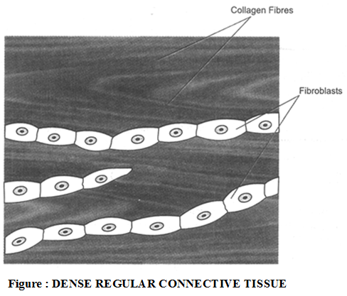 (a) Tendon : Tendons are cord-like, very tough, inelastic bundles of white collagen fibres bound together by areolar tissue. The cells present in the tendons are elongated fibroblasts which lie in almost continuous rows here and there. The tendons connect the skeletal muscles with the bones.
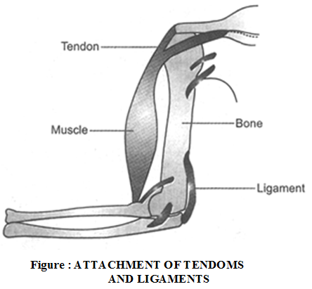 (b) Ligaments : Ligaments are cords formed by yellow elastic tissue in which many collagen fibres are bound together by areolar tissue. The fibroblasts are irregularly scattered. This tissue combines strength with great flexibility. The ligaments serve to bind the bones together.
Difference between tendon and ligament :
Tendons | Ligaments |
| 1. Tendons are very tough and inelastic. | 1. Ligaments are elastic. |
| 2. They connect the skeletal muscles with the bones. | 2. Ligaments connect bones to other bones at joints. |
| 3. Tendons are made up of white fibrous tissue. Yellow elastic fibres are, however, absent. | 3. Ligaments are made up of yellow elastic fibres. The white fibres also occur but they are very fine. |
| 4. Fibroblasts occur in rows. | 4. Fibroblasts lies scattered. |
3. Adipose tissue :
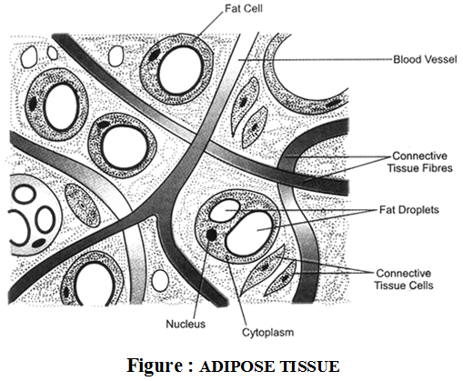
Adipose is primarily a fat storing tissue in which the matrix is packed with large, spherical or oval fat cells (or adipocytes). Each fat cell contains a large fat globule. The matrix also contains fibroblasts, macrophages, collagen fibres and elastic fibres. The adipose tissue is arranged in lobules encased in areolar tissue.
The adipose tissue is found beneath the skin, in the covering of the heart, around the blood vessels and kidney and in yellow bone marrow. This tissue stores fat and insulates the body against heat loss. It forms a shock aborbing cushion around the kidneys and the eyeballs. Bulbber in whales is, in fact, an insulating fat body. Similarly, hump in camel is also rich in adipose tissue.
4. Skeletal tissue :
Skeletal tissue forms the rigid skeleton which supports the vertebrate body, helps in locomotion and provides protection to many vital organs. There are two types of skeletal tissues.
- Cartilage
- Bone
 1. Cartilage
Characteristics. Cartilage is a hard but flexible skeletal tissue consisting of living cells embedded in a matrix. The cells (chondroblasts) become chondrocytes when get surrounded within special fluid-filled chambers, called lacunae (sing. lacuna). The lacunae (containing chondrocytes)are separated by the amorphous matrix (chondrin) that contains glycoproteins, collagen and elastic fibres. The surface of cartilage is surrounded by irregular connective tissue forming the perichondrium. Growth of cartilage occurs continuously due to multiplication of chondrocytes by mitosis, deposition of matrix within existing cartilage and from activity of the deeper cells of the perichondrium. Blood vessels and nerves are absent in the matrix.
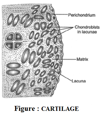
Occurrence. This tissue occurs in very few parts of the body. In humans, the cartilage occurs at the ends of long bones, the pinnae of ears, the ends of nose, in the walls of respiratory ducts, within intervertebral discs, etc. In sharks and rays, the entire skeleton is cartilage.
Functions. Cartilage is more compressible than bone. It absorbs stresses and provides flexibility to the body parts.
2. Bone
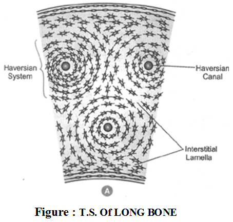
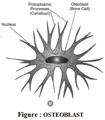
Characterisitics. Bone is a very strong and non-flexible vertebrate connective tissue. A compact bone consists of living bone cells. Called osteoblasts, embedded in a firm, calcified matrix. The osteoblasts are contained in lacunae (spaces) which are arranged in concentric circles present throughout the matrix. The lacunae are also traversed by nerves and blood vessels. The blood vessels passing through them provide nutrients to osteoblasts and help exchange of materials. The matrix in cmposed of about 30% organic materials (chiefly collagen fibres and glycoproteins) and 70% inorganic bone salts (mainly phosphates and charbonates of calcium and magnesium, hydroxyapatite, etc.). These inorganic salts are responsible for hardness of the bone.
Cartilage | Bone |
1. Cartilage is soft, elastic | 1. Bone is hard, tough and inelastic. |
2. Matrix of cartilage | 2. Matrix of bone is |
| 3. Cartilage do not have blood supply (except in perichondrium). | 3. Bones have rich |
| 4. Growth of cartilage is unidirectional. | 4. Growth of bone is |
Functions. Bones form endoskeleton of vertebrates. They provide levers for movement and support for soft parts of the body. Bones also protect many delicate tissues and organs.
4. Fluid Connective Tissue : (Vascular Tissue)
Fluid connective tissue links the different parts of body and maintains a continuity in the body. It includes blood and lymph.
1. Blood :
It is a fluid connective tissue.
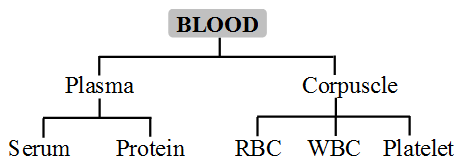 Functions of blood :
- Blood transports nutrients, hormones and vitamins to the tissues and transports excretory products from the tissues to the liver and kidney.
- The red blood corpuscles (RBC’s) carry oxygen to the tissues for the oxidation of food stuff.
- The white blood cells (WBC’s) fight disease either by engulfing and destroying foreign
bodies or by producing antitoxins and antibodies that neutrophils and harmful effects of germs. - Granulocytes include neutorphils, eosinophils and basophils.
- Agranulocytes include lymphocytes and monocytes.
- Blood platelets disintegrate at the site of injury and help in the clotting of blood.
2. Lymph:
Lymph is a colourless fluid that has filtered out of the blood capillaries. Red blood corpuscles and some blood proteins are absent in it. In the lymph, white blood cells are found in abundance.
Functions :
- Lymph transports the nutrients (oxygen, glucose) that may have filtered out of the blood capillaries back into the heart to be recirculated in the body.
- It brings CO2 and nitrogenous wastes from tissue fluid to blood.
