In this article, we are going to discuss the B.E. Biomedical Engineering, semester VI, Anna University connected to the regulation 2021 subject syllabus. Let’s see what’s more…
We tried our best to provide the following unit-wise BM3652 – Medical Image Processing detailed Syllabus. We sum up the appropriate textbooks and references to this page. If you have any doubts regarding the syllabus, you can simply comment below on the following page. Hope this information is useful. Share it with your classmates. Thanks for landing on this page.
If you want to know more about the B.E. Biomedical Engineering Syllabus connected to an affiliated institution’s four-year undergraduate degree program. We provide you with a detailed Year-wise, semester-wise, and Subject-wise syllabus in the following link B.E. Biomedical Engineering Syllabus Regulation 2021 Anna University.
Aim of Concept:
The objective of this course is to enable the student to
- Learn the fundamental concepts of medical Image Processing techniques.
- Understand the concepts of various image intensity transformation and filtering operations.
- Be familiar in the techniques of segmentation and restoration of medical images.
- Gain knowledge in medical image registration and visualization.
- Be familiar with the application of medical image analysis.
BM3652 – Medical Image Processing Syllabus
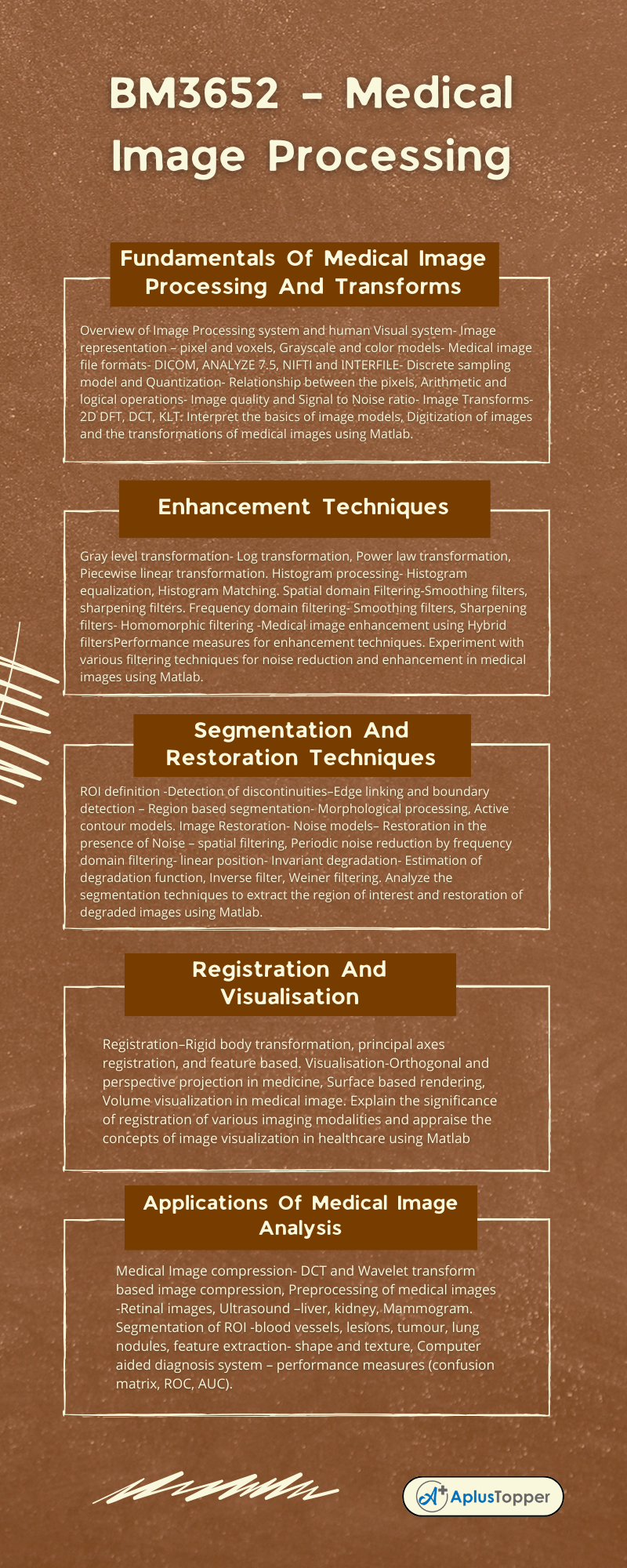
Unit I: Fundamentals Of Medical Image Processing And Transforms
Overview of Image Processing system and human Visual system- Image representation – pixel and voxels, Grayscale and color models- Medical image file formats- DICOM, ANALYZE 7.5, NIFTI and INTERFILE- Discrete sampling model and Quantization- Relationship between the pixels, Arithmetic and logical operations- Image quality and Signal to Noise ratio- Image Transforms- 2D DFT, DCT, KLT. Interpret the basics of image models, Digitization of images and the transformations of medical images using Matlab.
Unit II: Enhancement Techniques
Gray level transformation- Log transformation, Power law transformation, Piecewise linear transformation. Histogram processing- Histogram equalization, Histogram Matching. Spatial domain Filtering-Smoothing filters, sharpening filters. Frequency domain filtering- Smoothing filters, Sharpening filters- Homomorphic filtering -Medical image enhancement using Hybrid filtersPerformance measures for enhancement techniques. Experiment with various filtering techniques for noise reduction and enhancement in medical images using Matlab.
Unit III: Segmentation And Restoration Techniques
ROI definition -Detection of discontinuities–Edge linking and boundary detection – Region based segmentation- Morphological processing, Active contour models. Image Restoration- Noise models– Restoration in the presence of Noise – spatial filtering, Periodic noise reduction by frequency domain filtering- linear position- Invariant degradation- Estimation of degradation function, Inverse filter, Weiner filtering. Analyze the segmentation techniques to extract the region of interest and restoration of degraded images using Matlab.
Unit IV: Registration And Visualisation
Registration–Rigid body transformation, principal axes registration, and feature based. Visualisation-Orthogonal and perspective projection in medicine, Surface based rendering, Volume visualization in medical image. Explain the significance of registration of various imaging modalities and appraise the concepts of image visualization in healthcare using Matlab

Unit V: Applications Of Medical Image Analysis
Medical Image compression- DCT and Wavelet transform based image compression, Preprocessing of medical images -Retinal images, Ultrasound –liver, kidney, Mammogram. Segmentation of ROI -blood vessels, lesions, tumour, lung nodules, feature extraction- shape and texture, Computer aided diagnosis system – performance measures (confusion matrix, ROC, AUC).
Text Books:
- Rafael C. Gonzalez and Richard E. Woods, Digital Image Processing, Pearson Education, 3rd edition, 2016.
- Isaac N. Bankman, Handbook of Medical Image Processing and Analysis, 2nd Edition, Elsevier, 2009.
- Wolfgang Birkfellner, Applied medical Image Processing: A Basic course, CRC Press, 2011
References:
- Atam P.Dhawan, Medical Image Analysis, Wiley-Interscience Publication, NJ, USA 2003
- Rangaraj M. “Rangayyan, Biomedical Image Analysis”, 1st Edition, CRC Press, Published December 30, 2004.
- Joseph V.Hajnal, Derek L.G.Hill, David J Hawkes, “Medical image registration”, Biomedical Engineering series, CRC Press,2001
- Milan Sonka, Image Processing, Analysis And Machine Vision, Brookes/Cole, Vikas Publishing House, 2nd edition, 1999.
- Anil Jain K, Fundamentals of Digital Image Processing, PHI Learning Pvt. Ltd., 2011.
Related Posts On Semester – VI:
Must Read:
